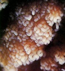
| Seminiferous tubule. The highly coiled seminiferous tubules within the testes are lined with germinal epithelium. Spermatogenesis occurs in the germinal epithelium. Photo source: Nilsson, Lennart 1990. A Child is Born. Delacorte Press/Seymour Lawrence. ISBN 0-385-30237-1 (Page 27) |
 |
| Ovary. The uterus is on the left side and an ovarian ligament extends to the right holding the bulging ovary. At the far left is the infundibulum of the fallopian tube (also called the uterine tube or oviduct) surrounded by fimbriae. Photo source: Nilsson, Lennart 1990. A Child is Born. Delacorte Press/Seymour Lawrence. ISBN 0-385-30237-1 (Page 19) |
 |
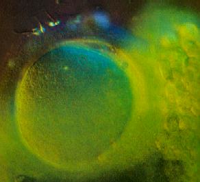
| Fertilization. A human oocyte with several sperm embedded in the outer portion of the zona pellucida. A few cells of the corona radiata can be seen. Fertilization usually occurs in the ampulla of the fallopian tube. Photo source: Nilsson, Lennart 1990. A Child is Born. Delacorte Press/Seymour Lawrence. ISBN 0-385-30237-1 (Page 47) |
 |
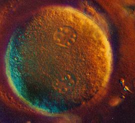
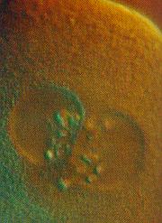
| Pronuclei. A fertilized human oocyte with two pronuclei. One pronucleus contains the oocyte genetic material (the oocyte has completed meiosis II), the other contain the genetic material contributed by the sperm. A few sperm that did not fertilize this oocyte are embedded in the outer portion of the zona pellucida. Photo source: Nilsson, Lennart 1990. A Child is Born. Delacorte Press/Seymour Lawrence. ISBN 0-385-30237-1 (Page 56) |
 |
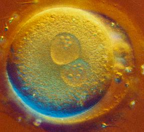
| Pronuclei. A fertilized human oocyte with two pronuclei that have nearly joined. One pronucleus contains the oocyte genetic material (the oocyte has completed meiosis II), the other contain the genetic material contributed by the sperm. The material on the right side, within the zona pellucida, may be polar bodies. A few sperm that did not fertilize this oocyte can be seen outside the zona pellucida. Photo source: Nilsson, Lennart 1990. A Child is Born. Delacorte Press/Seymour Lawrence. ISBN 0-385-30237-1 (Page 56) |
 |
| Pronuclei. A fertilized human oocyte with two pronuclei that are fusing. As the nuclear envelopes of the pronuclei break down, the two haploid sets of chromosomes mingle and the zygote is formed. Photo source: Nilsson, Lennart 1990. A Child is Born. Delacorte Press/Seymour Lawrence. ISBN 0-385-30237-1 (Page 56) |
 |
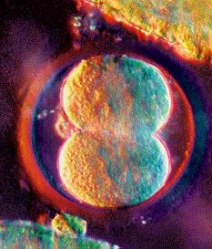
| Two-cell. A human embryo about 30 hours after fertilization. The first cleavage seems to be complete. The surrounding circular structure is the zona pellucida. Photo source: Nilsson, Lennart 1990. A Child is Born. Delacorte Press/Seymour Lawrence. ISBN 0-385-30237-1 (Page 50) |
 |
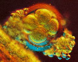
| Eight-cell. A human embryo about two days after fertilization. Four blastomeres are visible and there are probably four more beneath those. The zona pellucida and some surrounding corona radiata cells can be seen. Photo source: Nilsson, Lennart 1990. A Child is Born. Delacorte Press/Seymour Lawrence. ISBN 0-385-30237-1 (Page 60) |
 |
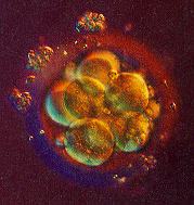
| 16-cell. A human embryo about 2.5 days after fertilization. The zona pellucida and some surrounding corona radiata cells can be seen. Photo source: Nilsson, Lennart 1990. A Child is Born. Delacorte Press/Seymour Lawrence. ISBN 0-385-30237-1 (Page 60) |
 |
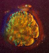
| Morula. A human embryo about 2.5-3 days after fertilization. The term morula is used starting at 12-16 cells until the blastocyst stage beginning about 50 cells. We can tell the morula is not much larger than the zygote because the zona pellucida is still present. Photo source: Nilsson, Lennart 1990. A Child is Born. Delacorte Press/Seymour Lawrence. ISBN 0-385-30237-1 (Page 61) |
 |
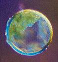
| Blastocyst. Human embryos about 4-5 days after fertilization. The term blastocyst (or blastula) is used when there are about 50 cells and the blastocyst cavity (blastocoel) begins to form. The zona pellucida degenerates as the blastocyst grows (but is still present here). Photo source: Nilsson, Lennart 1990. A Child is Born. Delacorte Press/Seymour Lawrence. ISBN 0-385-30237-1 (Page 62) |
  |
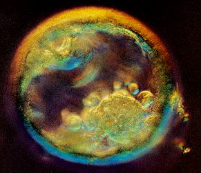
| Blastocyst. A human embryo about 5 days after fertilization. The term blastocyst or blastula is used when there are about 50 cells and the blastocyst cavity (blastocoel) begins to form. The inner cell mass (ICM) is located at the lower right. Photo source: Nilsson, Lennart 1990. A Child is Born. Delacorte Press/Seymour Lawrence. ISBN 0-385-30237-1 (Page 63) |
 |
| 3-4 Weeks. A human embryo early in the fourth week after fertilization (about 22-23 days old and 2-3 mm long). The neural tube is forming (neurulation). The embryo's cranial aspect is at the upper right. The neural folds are closing over along the mid-dorsal line. The neural folds at the rostral 1/3 of the tube are especially thick. Photo source: Nilsson, Lennart 1990. A Child is Born. Delacorte Press/Seymour Lawrence. ISBN 0-385-30237-1 (Page 76) |
 |
| 4 Weeks. A human embryo in the fourth week after fertilization (about 26-27 days old and 4-5 mm long). The ventricle of the heart is clearly visible. The well developed umbilical vessels connect the embryo's circulation to the placenta. Three pairs of branchial arches have formed. Photo source: Nilsson, Lennart 1990. A Child is Born. Delacorte Press/Seymour Lawrence. ISBN 0-385-30237-1 (Page 79) |
 |
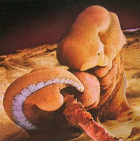
| 4 Weeks. A human embryo late in the fourth week after fertilization (about 28-32 days old and 5-6 mm long). The lower limb buds are well formed and the embryo's curving tail is prominent. This S.E.M. has been colored showing the location of the somites. Photo source: Nilsson, Lennart 1990. A Child is Born. Delacorte Press/Seymour Lawrence. ISBN 0-385-30237-1 (Page 80) |
 |
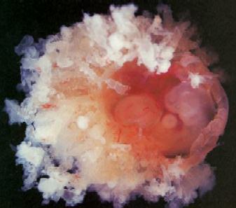
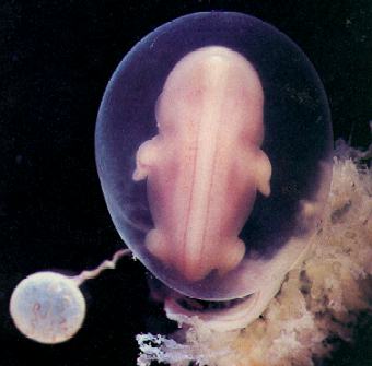
| 5 Weeks. A human embryo in the fifth week after fertilization (about 32-36 days old and 6-7 mm long). Pale chorionic villi surround the amniotic cavity. The membranes have been cut open to reveal the embryo. The embryo's head is at the far right. Photo source: Nilsson, Lennart 1990. A Child is Born. Delacorte Press/Seymour Lawrence. ISBN 0-385-30237-1 (Page 80) |
 |
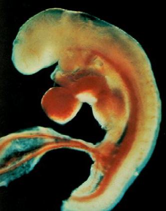
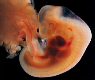
| 6 Weeks. A human embryo in the sixth week after fertilization (about 38-40 days old and 10-11 mm long). Pigment can be seen in the retina. The cerebral vesicles are well developed. The pontine flexure is clearly visible (the roof of the metencephalon is so thin it seems transparent). Photo source: Nilsson, Lennart 1990. A Child is Born. Delacorte Press/Seymour Lawrence. ISBN 0-385-30237-1 (Page 81) |
 |
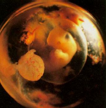
| 6 Weeks. A human embryo late in the sixth week after fertilization (about 41-43 days old and 11-14 mm long). Digital rays can be seen in the hand plates. The amnion surrounds the entire embryo. Blood vessels can be seen in the wall of the yolk sac. Photo source: Nilsson, Lennart 1990. A Child is Born. Delacorte Press/Seymour Lawrence. ISBN 0-385-30237-1 (Page 83) |
 |
| 6 Weeks. A human embryo in the sixth week after fertilization (about 41 days old and 11-14 mm long). We are looking at the developing face. The stomodeum (mouth opening) can be seen between the maxillary and mandibular processes. The nasal pits have formed nostrils. The labial grooves have closed. The medial nasal elevations are beginning to merge. Photo source: Nilsson, Lennart 1990. A Child is Born. Delacorte Press/Seymour Lawrence. ISBN 0-385-30237-1 (Page 83) |
 |
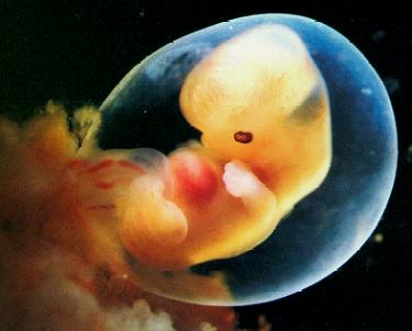
| 6 Weeks. A human embryo in the sixth week after fertilization (about 41-43 days old and 11-15 mm long). The mid-dorsal line shows the neural tube. Cranially, the embryo's head is bent ventrally at the cervical flexure. The amnion and yolk sac are clearly seen. Photo source: Nilsson, Lennart 1990. A Child is Born. Delacorte Press/Seymour Lawrence. ISBN 0-385-30237-1 (Page 84) |
 |
| 7 Weeks. A human embryo early in the seventh week after fertilization (about 43-46 days old and 14-17 mm long). Notches between the digital rays can be seen. Photo source: Nilsson, Lennart 1990. A Child is Born. Delacorte Press/Seymour Lawrence. ISBN 0-385-30237-1 (Page 84) |
 |